


Hip fusion arthrosis (deformation of arthrosis, coxarthrosis, osteoarthrosis) is a slowly progressive degenerative-degenerative disease, leading to destroying the affected joint, persistent pain and restriction of mobility.
The disease affects people over 40, women get sick several times more often than men.
In the overall structure of arthrosis, hip joint arthrosis belongs to the main role.This is due to the widespread congenital pathology of the hip joints (dysplasia), as well as important physical exercise, for which these joints are sensitive to.
Risk factors and causes of hip arthrosis arthrosis
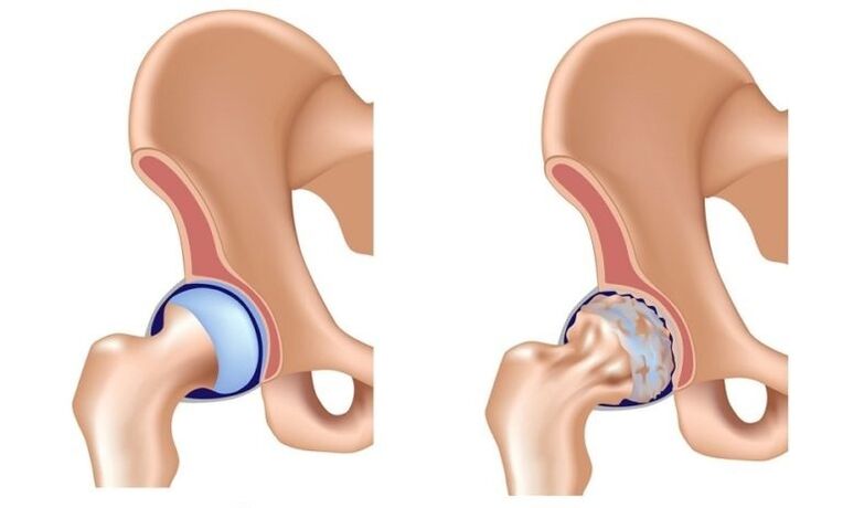
In the pathological mechanism of developing arthrosis of the hip joint, the main role belongs to a change in the physico-chemical characteristics of the synovial fluid (intra-articular), as a result of which becomes denser and viscous.This exacerbates its lubricating qualities.When moving, the surfaces of the joint cartilage begin to rub against each other, become rough, covered with cracks.Small particles of the hyaline cartilage are abandoned and fall into the articular cavity, causing the development of aseptic inflammation in it.As the disease progresses in the inflammatory process, bone tissue is attracted to the inflammatory process, which leads to aseptic necrosis of the femoral head and surface of the acetabulum, osteophyte formation (bone growth), increasing inflammation and causing severe pain during movement.
In the late extension of the arthrosis of the hip joint, inflammation is also thrown into the surrounding tissue joint (blood vessels, nerves, ligaments, muscles), which leads to the appearance of periartrit signs.As a result, the hip knot is completely destroyed, its functions are lost, the movement in it ceases.This condition is called ankylosis.
Causes of hip union arthrosis:
- the congenital edge of the thighs;
- hip dysplasia;
- aseptic necrosis of the femur's head;
- Peter disease;
- Hip joint damage;
- infectious arthritis of the hip joint;
- gonartrosis (deformity of the knee joint osteoarthrosis);
- osteochondrosis;
- Excess weight;
- professional sports;
- flat feet;
- curvature of the spine;
- A sedentary lifestyle.
Pathology is not inherited, but the child inherits the features of the structure of the skeletal muscle system from his parents, which can cause arthrosis of the hip joint under these conditions.This explains the fact of the existence of families whose incidence is higher than in the general population.
Forms of illness
Depending on the etiology, the arthrosis of the hip joint is divided into elementary and secondary.Secondary arthrosis develops against the backdrop of other diseases of the hip joint or its damage.The primary form is not about the previous pathology, the reason for its development is often not possible, in this case they talk about idiopathic arthrosis.
Coxartrosis is one or bilateral.
PHASE
Three stages (stairs) are distinguished during hip arthrosis arthritis:
- Initial - pathological changes are expressed little, provided that timely and adequate treatment is reversible.
- Progressive coxarthrosis - characterized by a gradual increase in symptoms (pain in the joint and damaged its movement), changes in joint tissue are already irreversible, but therapy may slow down degenerative processes.
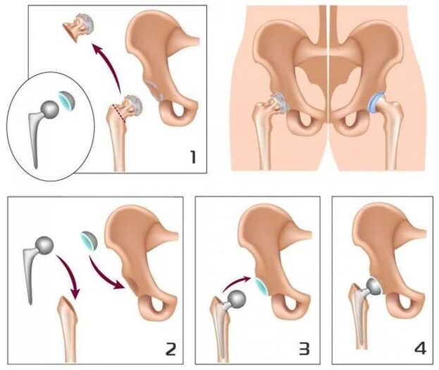
- The final movement in the union is lost, ankylosis is formed.Treatment is only surgically possible (replacing the fusion with artificial).
Endoprosthetic operations in 95% of cases provide a complete restoration of limb mobility, restore patient performance.
Symptoms of hip joint arthrosis
The main signs of hip union arthrosis:
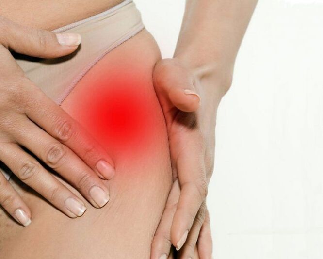
- pain in the groin, hips and knees;
- a feeling of stiffness in the affected joint and limiting its mobility;
- madness;
- restriction of abduction;
- Atrophic changes in the thigh muscles.
The presence of some symptoms of arthrosis of the hip joint, as well as their severity depends on the degree of the disease.
At the 1st degree of hip arthrosis, patients complain of pain in the affected joint that occurs under the influence of physical activity (prolonged walking, jogging).In some cases, the pain is located in the knee or thigh area.After a short break, the pain passes on its own.The volume of limb movements is fully preserved, the walking does not break down.The following changes are listed in radiographs:
- small unequal landing on the lumen of the common gap;
- Osteophytes located at the inner edge of the bend.
Anydo change from the neck and the femoral head is not detected.
With the II degree of hip joint arthrosis, the pain also appears at rest, including at night.After physical activity, the patient begins to lame, a characteristic "duck" walking is formed.Thus, which starts start pain - after a long period of immobility, the first steps cause pain and discomfort, which then pass and then return after a long load.At the affected node, the volume of movements (abduction, internal rotation) is limited.Radiography shows that the common gap is unevenly narrowed and its lumen is 50% of the rate.Osteophytes are located both along the inner and outer edge of the joint cavity, going beyond the borders of the cartilage.The femoral head contours become uneven due to deformation.
With degree III of arthrosis of the pain hip, intense and constant hip joint, not stopping at night.Walking is significantly difficult, the patient is forced to rely on the cane.The volume of movements in the affected joint is severely limited, later stops completely.Due to the atrophy of the hip muscle, the pelvis deviates on the frontal plane and the limbs are cut.Trying to make up for this abbreviation, patients when walking are forced to reject the body towards the lesion, which further increases the load on the injured joint.In radiographs, numerous bone increases, a significant narrowing of the joint gap and a marked increase in the femur's head are detected.
Troubleshooting
The diagnosis of the arthrosis of the hip node is based on the data of the clinical figure of the disease, the results of medical examination and instrumental studies, among which the main value belongs to the methods of visualization - radiography, calculated or magnetic resonance image.They allow not only to determine the presence of hip joint arthrosis and evaluate its rank, but also identify the potential cause of the disease (trauma, youth epiphysiolis, Peter disease).
Differential diagnosis of hip joint arthrosis with other diseases of the skeletal muscle system is quite complicated.In degree II and III of the arthrosis of the hip joint, muscle atrophy develops, which can cause intense pain in the characteristic knee or gonarthrosis (knee disease).For the differential diagnosis of these states, palpation of the knee and hip joints is palpable, the volume of movement in them is determined, and they are also examined by radiologically.
In spinal diseases, in some cases, the nerve roots of the spinal cord with the development of pain syndrome are squeezed.Pain can radiate to the hip joint area and imitate the clinical appearance of its loss.However, the nature of the pain with radical syndrome is a little different than with the arthrosis of the hip joint:
- Pain occurs as a result of lifting weight or sharp movement difficult, not under the influence of physical exercise;
- The pain is localized in the gluteal, not in the inguinal region.
With radicular syndrome, the patient can calmly take the leg to the side, while with arthrosis of the hip joint, the abduction is limited.A characteristic sign of radicular syndrome is a positive symptom of tension - the appearance of a sharp pain when you try to raise a straight foot on the patient's back.
Hip arthritis affects people over 40, women get sick several times more often than men.
Hip joint arthrosis should also be differentiated with loyal bursitis (trochanteritis).The voluntary bursitis develops faster, within a few weeks.It is usually preceded by an important physical exercise or injury.In this disease, the pain is much more pronounced than with the arthrosis of the hip joint.At the same time, the shortening of the limbs and the restriction of its mobility has not been detected.
Clinical photography of reactive atypical arthritis and ankylosing spondylitis may resemble the clinical manifestations of hip joint arthrosis.However, pain occurs in patients mainly at night or at rest, when walking does not intensify, but, on the contrary, weakens.In the morning, patients mark rigidity in the joints, which passes after a few hours.
Hip joint arthrosis treatment
Orthopedians are engaged in the treatment of hip joint arthrosis.With the degree I and II of the disease, conservative therapy is indicated.With pronounced pain syndrome, patients are prescribed non -steroidal anti -inflammatory drugs in a short course.They should not be accepted for a long time, as they are not only able to have a negative effect on the organs of the gastrointestinal tract, but also suppress the regenerative abilities of the hyaline cartilage.
In the hip joint arthrosis treatment regimen, they include chondroprotectors and vasodilators, which creates optimal opportunities for resetting damaged cartilage tissue.With pronounced muscle spasm, it can require the appointment of central muscle relaxants.
In those cases where it is not possible to stop the pain syndrome with non -steroidal anti -inflammatory drugs, using intraarticular corticosteroid injections.
Local treatment of hip joint arthrosis using heat ointments allows you to reduce muscle spasm and somewhat weaken pain due to distractional action.
In the complex therapy of the hip joint arthrosis, physiotherapeutic methods are also used:
- magnetotherapy;
- inductootermia;
- UHF;
- Laser therapy;
- ultrasound treatment;
- massage;
- Medical gymnastics;
- Manual therapy.
Dietary nutrition for hip joint arthrosis is aimed at correcting body weight and normalizing metabolic processes.Lowering body weight reduces the load on the hip joints and thus slows down the progression of the disease.
To discharge the affected joint, the doctor may recommend that patients go to crutches or cane.
With degree III of the hip joint arthrosis, conservative treatment is inefficient.In this case, it is possible to improve the patient's condition, it is possible to restore normal mobility only as a result of surgical intervention - replacing the artificial (joint endoprosthetic endoprosthetic) node.
Possible consequences and complications
The most serious complication of progressive arthrosis of the hip joint is disability due to loss of movement in the joint.With bilateral coksartrosis, the patient loses his ability to move independently and needs constant external care.A long bed stay in one position creates the preconditions for the emergence of stagnant (hypostatic) pneumonia, which is difficult to deliver and can lead to death.
The pathology is not inherited, but the child inherits the features of the structure of the musculoskeletal system from his parents, which can cause arthrosis of the hip joint.
Predict
Hip arthritis is a chronic progressive disease, which can only be cured in the early stages, undergoing elimination of the cause of the disease.In other cases, therapy allows you to slow its flow, however, over time, there is a need for implantation of hip endoprostese.Such operations in 95% of cases provide a complete restoration of limb mobility, restore patient performance.The service life of modern prosthesis is 15-20 years, after which they are subject to replacement.
PREVENTION
Prevention of hip joint arthrosis is aimed at eliminating the causes that can lead to the development of this disease, and includes:
- timely detection and treatment of diseases and damage to the hip joint;
- Rejecting a sedentary lifestyle, regular physical activity, but not redundant;
- body weight control;
- rational food;
- Rejecting bad habits.